When you intend to move your body voluntary, motor commands generated inside your brain are transmitted from the motor cortex to the spinal cord through nerve fibers. In the spinal cord, the motor commands are integrated and coordinated with the feedback signals from muscles, skin and so forth, and sent to the motor neurons which are connected to the muscle fibers.
When motor neurons are activated, all of the muscle fibers innervated by the neuron contract and produce force that causes joint movement.
Any trouble in the process from the cerebral nervous system to the musculo-skeletal system may develop motor dysfunction. Parkinson's disease is a neurological brain disorder that results from degeneration of neurons in the basal ganglia which controls movement. The degeneration of neurons creates a lack of the neurotransmitter called “dopamine”, causing the movement dysfunctions. The motor symptoms of Parkinson's disease include resting tremor, rigidity, bradykinesia and postural instability, but the mechanism whereby such symptoms are generated remains unexplained. We are developing an integrated simulator of the central nervous and musculo-skeletal systems of the entire human body with an aim to provide a useful tool for investigating the mechanisms underlying motor dysfunctions, and exploring effective therapeutic approaches through the use of computer simulations.
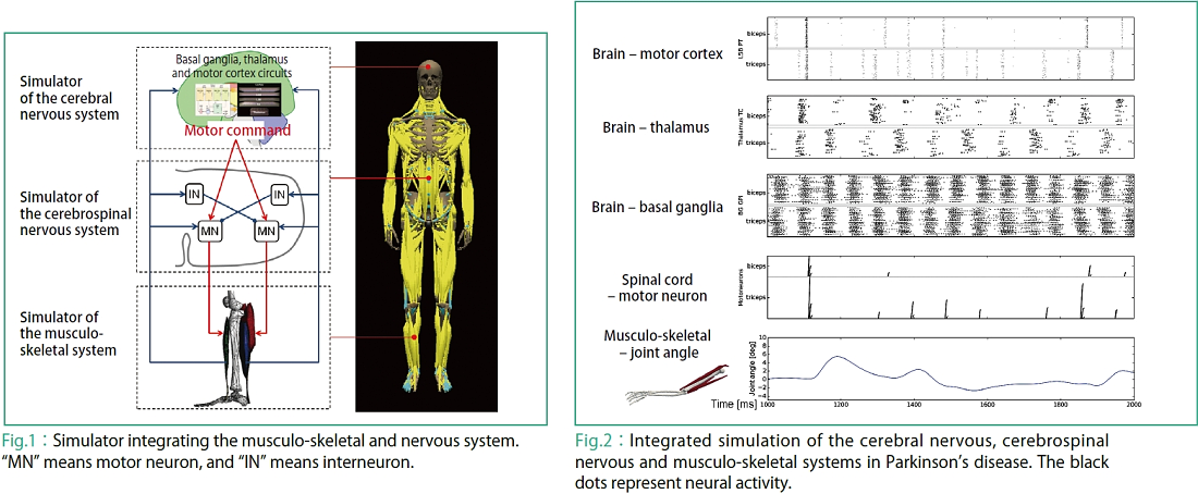
Our simulator is an integration of a simulator of the cerebral nervous system consisting of the basal ganglia, thalamus and motor cortex circuits, a simulator of the cerebrospinal nervous system computing the activity of motor neurons, and a simulator of the musculo-skeletal system computing behaviors of the musculo-skeletal system from the activity of motor neurons, which were developed by the Doya Research Group (Okinawa Institute of Science and Technology Graduate University), the Nakamura Research Group (The University of Tokyo), and the Takagi Research Group (The University of Tokyo) respectively, so as to expand them over the entire human musculo-skeletal system (Fig. 1).
Fig. 2 shows an example of an integrated simulation based on a central nervous system model and a human upper arm musculo-skeletal model in Parkinson's disease. The neural activities in the motor cortex, thalamus, basal ganglia in the brain, the activity of motor neurons in the spinal cord and the joint angle of an elbow joint are shown through time. The upper side of each neuron is projected to the biceps muscle, while its lower side is projected to the triceps muscle. In the basal ganglia, abnormal neural activity specific to patients with Parkinson's disease are reproduced. For neurons in the thalamus, alternate neural activities are reproduced with a frequency similar to that of Parkinsonian tremors (4 to 6Hz) in the biceps and triceps. The output from the simulator of the cerebral nervous system is sent to the spinal cord as the pyramidal tract activity of the motor cortex, and motor neuron activity is computed by the simulator of the cerebrospinal nervous system. The simulator of the musculo-skeletal system computes the force of contraction based on motor neuron activity, and tremor-like movements are observed in the elbow joint.
The development of this integrated simulator enables the simulation of human body movements originating from activity of the central nervous system. Using this simulator, we will proceed further with our academic studies with an aim to reproduce motor symptoms such as Parkinsonian tremor and rigidity, understand the mechanism generating such symptoms, and eventually develop treatment methods.
Integrated simulations of the nervous system and musculo-skeletal system for reproducing Parkinson’s disease symptoms
|
![]()
ZOOM IN Theme 4 From supercomputing analysis of big cancer genome data to life science and medical care

 |
 |
 |
 |
 |
|---|